PFO/ASD Closure
What is a PFO and an ASD?
Patent Foramen Ovale (PFO)
The heart is divided into four chambers. The upper chambers are called the right and left atria. The lower chambers are the right and left ventricles. In fetal circulation the foramen ovale is an opening that allows blood to bypass the lungs and go directly from the right atria to the left atria. Shortly after birth the higher pressure in the left atria and the lower pressure in the right atria causes permanent closure of the foramen ovale in the majority of people. A PFO occurs when the opening does not close. This opening can allow blood to pass from the right atria to the left atria. Many times a PFO is not discovered until adulthood. PFO’s are suspected to be a cause of cryptogenic stroke (a stroke that cannot be linked to a specific cause). Research suggests there may be a link between PFO’s and migraine headaches.
Atrial septal defect (ASD)
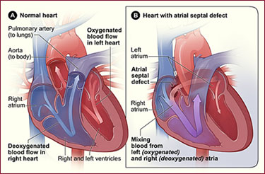
An ASD is a hole in the part of the septum that separates the atria—the upper chambers of the heart. This heart defect allows oxygen-rich blood from the left atrium to flow into the right atrium instead of flowing to the left ventricle as it should. Many children who have ASDs have few, if any, symptoms.

Figure A shows the structure and blood flow in the interior of a normal heart. Figure B shows a heart with an atrial septal defect, which allows oxygen-rich blood from the left atrium to mix with oxygen-poor blood from the right atrium.
An ASD can be small or large. Small ASDs allow only a little blood to leak from one atrium to the other. Very small ASDs don't affect the way the heart works and don't require any treatment. Many small ASDs close on their own as the heart grows during childhood.
Medium to large ASDs allow more blood to leak from one atrium to the other, and they're less likely to close on their own.
Half of all ASDs close on their own or are so small that no treatment is needed. Medium to large ASDs that need treatment can be repaired using a catheter procedure or open-heart surgery.
Catheter Procedures
Catheter procedures are much easier on patients than surgery because they involve only a needle puncture in the skin where the catheter (thin, flexible tube) is inserted into a vein or an artery.
Doctors don't have to surgically open the chest or operate directly on the heart to repair the defect(s). This means that recovery may be easier and quicker.
The use of catheter procedures has grown a lot in the past 20 years. They have become the preferred way to repair many simple heart defects, such as atrial septal defect (ASD).
For an ASD, the doctor inserts a catheter through a vein and threads it into the heart to the septum. The catheter has a tiny, umbrella-like device folded up inside it.
When the catheter reaches the septum, the device is pushed out of the catheter. It's positioned so that it plugs the hole between the atria. The device is secured in place and the catheter is then withdrawn from the body.
To help guide the catheter, doctors often use echocardiography (echo) or transesophageal (tranz-ih-sof-uh-JEE-ul) echocardiography (TEE) and angiography (an-jee-OG-ra-fee).
TEE is a special type of echo that takes pictures of the back of the heart through the esophagus (the passage leading from the mouth to the stomach). TEE also is often used to examine complex heart defects.
Source: National Heart, Lung, and Blood Institute (NHLBI)